

In microscopes, objective lenses are the most complex and most important part of the microscope. The objective lenses, which are designed as multi-element lenses located closest from the specimen, the function is to receive light emitted by the specimen, and perform the crucial task of producing real image, then get it received by the eyepiece or computers. They are used in various fields such as scientific research, biology, industry, and laboratory work.
Classifying Objective Lenses
A wide range of objective lenses is available for selection, depending on their design and quality. They can be roughly classified based on intended purpose; microscopy method; performance; magnification; aberration correction. In this article, we will introduce the five objective lenses classified by aberration correction.
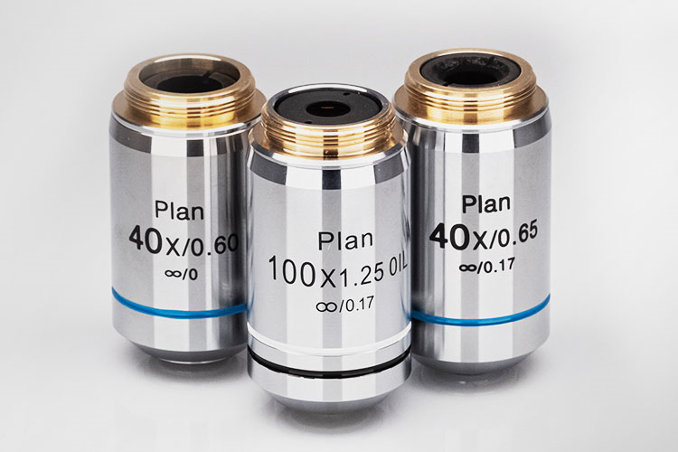
- Achromatic Objectives: These are the most affordable and most widely used lenses. If there are no markings stating that they are not achromatic, we can assume that they are. They correct chromatic aberration in red and blue light, so they can meet at one focal point, and spherical aberration for green light.
- Plan Achromatic Objectives (Achroplan): In a non-corrected objective, you can find sharp focus around the edges of the field of view or the center of the field of view. This type of microscope lens offers correction for chromatic aberration in two wavelengths and a correction of field curvature.
- Plan Fluorite Objectives (Plan Semi-Apochromats): These lenses were originally manufactured using the mineral fluorite, but now are mainly made of synthetic materials. These versatile lenses provide improved chromatic aberration correction and a flat field, with spherical aberration corrected for two wavelengths.
- Plan Apochromatic Objectives (Plan Apo): Compared to Plan Fluorite Objectives, these lenses have a better transmission in the 400nm to 100nm range. These are chromatically corrected for three colors (red, green, and blue), meaning that those three wavelengths of light could focus on one focal point. In addition, they also correct spherical aberration for two or three wavelengths.
- Super Apochromatic Objectives: These lenses deliver outstanding performance over the entire visible to near-infrared field of view with axial color aberration.
Key Specifications
- Aberration Correction or Resolution: Spherical and chromatic aberrations limit the resolution of conventional microscopes. Lenses with a high degree of aberration correction result in high-resolution images over the entire field of view.
- Magnification: A microscope’s ability to produce larger images is referred to as magnification. High magnification objectives provide extremely detailed images of specimens. The term magnification is often confused with the term resolution, which describes the ability of an imaging system to show detail in the object that is being imaged. While high magnification without high resolution may result in very small microbes visible, it will not allow observers to distinguish between microbes or sub-cellular parts of a microbe. Avantier has spent years designing and manufacturing to satisfy both magnification and resolution requirements simultaneously.
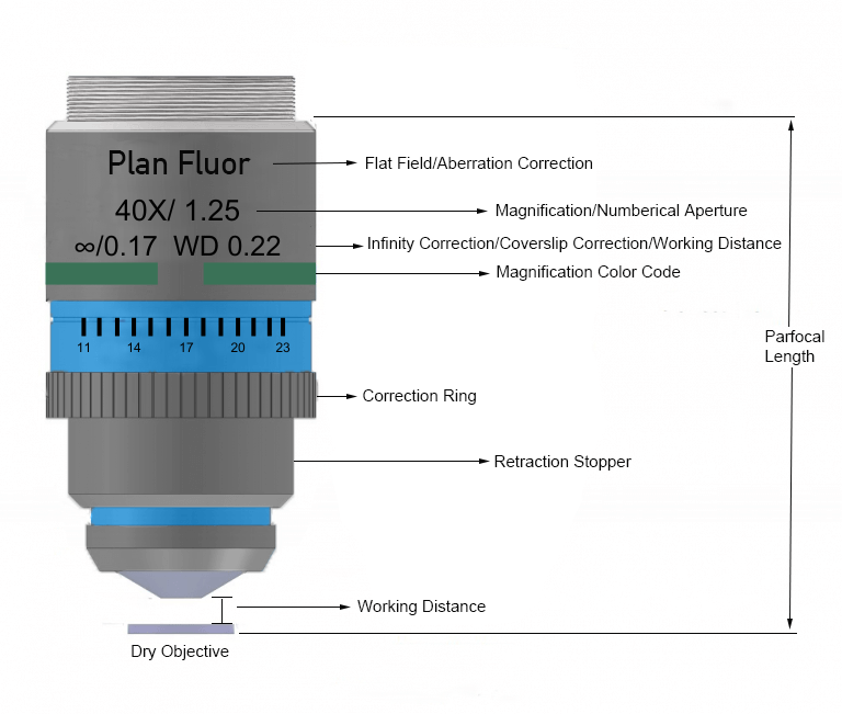
- Numerical Aperture (NA): NA is the measure of its capability to gather light and to resolve fine specimen details at a fixed object. A lens with a high NA collects more light and can resolve finer specimen details at a fixed distance.
- Conjugate Distance: Objectives are corrected for a specific projection distance. In finite conjugate optical design, light from a non-infinite source is focused down to a point. In infinity-corrected optical systems, light emitted from the specimen passes through the objective lens and enters the tube lens as an infinity parallel beam, forming a real image.
- Cover Glass: Objectives are usually corrected for a specific cover glass thickness, with 0.17 millimeters being the standard.
- Immersion Media: The main purpose of using different types of immersion media is to minimize the refractive index between the objective and the sample. It is crucial to use the correct media, such as water, oil, or air/dry, as specified by the objective.
- Working Distance: The distance between the front end of the microscope objective and the surface of the specimen at which the sharpest focus is achieved. Proper positioning is important to obtain a good image at the specified magnification.
- Parfocal Length: The distance from the shoulder of the objective to the sample plane.
- Working Wavelength(s): Objectives are corrected for specific wavelengths, with shorter wavelengths yielding higher resolution.